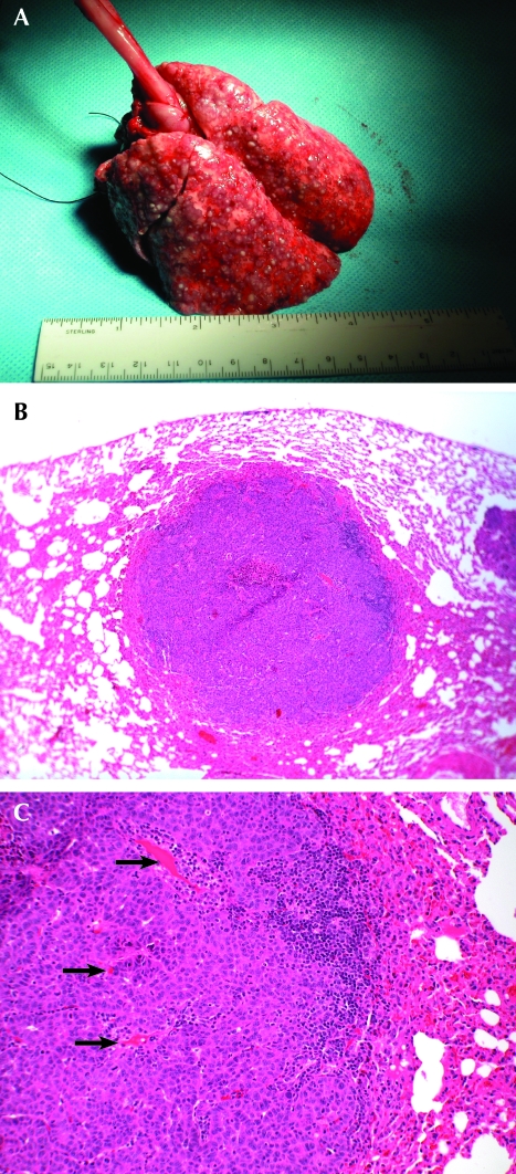
Figure 4.
(A) Hematogenous spread of VX2 tumors to the lungs. Note the cobblestone appearance of the normally smooth lungs. (B) Tumor that localized to the lung 20 d after IV implantation with previously frozen cells. Hematoxylin and eosin; magnification, ×40. (C) High magnification of the lung tumor. Hematoxylin and eosin; magnification, ×100. Arrows point to notable vascularization.

