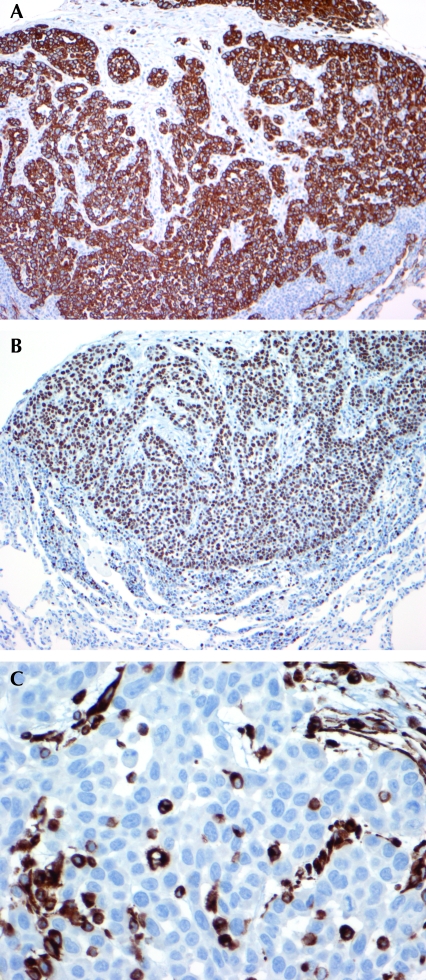
Figure 5.
(A) Immunohistochemistry for keratin AE1/AE3 shows strong positive cytoplasmic staining in the tumor cells. Immunoperoxidase stain; magnification, ×100. (B) Immunohistochemistry for the proliferation marker Ki67 is positive in 90% of the tumor cells. Immunoperoxidase stain; magnification, ×100. (C) Immunohistochemistry for vimentin shows scattered tumor cell positivity. Immunoperoxidase stain; magnification, ×400.

