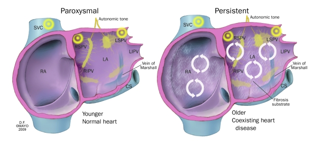
FIGURE 1.
Important differences between the paroxysmal and persistent forms of atrial fibrillation. These differences have implications for management and for outcome expectations. Circular arrows represent rotors. CS = coronary sinus; LA = left atrium; LIPV = left inferior pulmonary vein; LSPV = left superior pulmonary vein; RA = right atrium; RIPV = right inferior pulmonary vein; RSPV = right superior pulmonary vein; SVC = superior vena cava.

