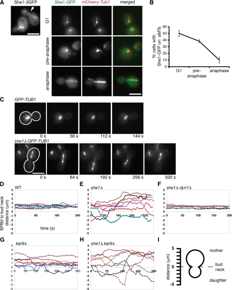
Figure 1.
She1p is a microtubule and bud neck–associated protein required to inhibit dynein activity before anaphase. (A) She1-3GFP localizes along the entire length of the mitotic spindle and at the bud neck (arrowhead). She1-GFP predominately localizes along astral microtubules in G1 and pre-anaphase cells, but not in anaphase cells. (B) Quantification of She1-GFP localization to aMTs. (C) Time-lapse images of pre-anaphase cells expressing GFP-Tub1. Cells were arrested with hydroxyurea to prevent entry into mitosis. Cell shape is outlined in white. Scale bar, 5 μm. (D through H) Graphs plotting the distances between the daughter-bound SPB (SPBd) and the bud neck over time for cells expressing GFP-Tub1. Each line represents the spindle position for an individual cell. Comparison of spindle position between wild-type (n = 9; D), she1Δ (n = 7; E), she1Δ dyn1Δ (n = 7; F), kar9Δ (n = 8; G), and she1Δ kar9Δ cells (n = 7; H). The addition of the dyn1Δ mutation, but not the kar9Δ mutation, eliminates spindle transiting seen in she1Δ cells. (I) Diagram depicting the measurements represented in D through H. Positive distances indicate that the SPBd was in the mother cell, whereas negative distances indicate that the SPBd was in the daughter cell.

