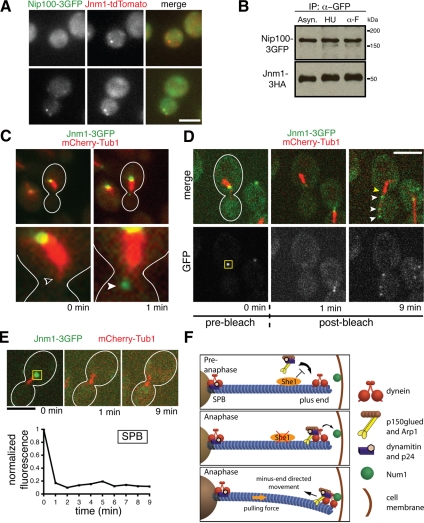
Figure 5.
Dynactin exists in complete and incomplete complexes that are spatially distinct. (A) Approximately 60% of Jnm1-tdtomato colocalizes with Nip100-3GFP in asynchronous cells (n = 114). (B) Immunoprecipitation of Nip100-3GFP from asynchronous (Asyn.), hydroxyurea-arrested (HU), and alpha factor–arrested (α-F) cells. Essentially equal amounts of Jnm1-3HA copurify with Nip100-3GFP at different cell cycle stages, indicating that a complete dynactin complex exists throughout the cell cycle. (C) Before anaphase, Jnm1-3GFP localizes to the SPB but is absent from the aMT plus end (unfilled arrowhead). Near the anaphase transition, Jnm1-3GFP suddenly appeared at the plus end (filled arrowhead), rather than being transported from the SPB. Anaphase onset was indicated by rapid elongation of the mitotic spindle shortly after Jnm1-3GFP appeared on the aMT (not shown). (D) Jnm1-3GFP fluorescence at the SPB was photobleached in pre-anaphase cells. The yellow box represents the area that was photobleached at t = 0.5 min. Appearance of GFP foci (white arrowheads) occurred along the aMT as the cell entered anaphase. There was no recovery of GFP fluorescence at the SPB (yellow arrowhead). (E) The GFP fluorescence at the SPB in a different cell also did not recover after photobleaching, indicating that SPB-localized dynactin subcomplexes are very static. The yellow box represents the area that was photobleached at t = 0.5 min. Scale bars, 5 μm. (F) A model for cell cycle control of dynein activation. She1p precludes stable association of the complete dynactin complex with dynein until anaphase.

