Figure 1.
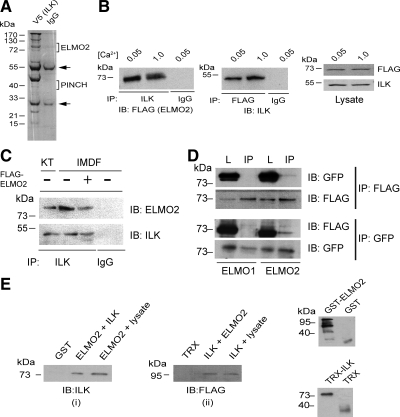
Interaction of ILK with ELMO2. (A) Lysates prepared from primary cultured human keratinocytes infected with a V5-tagged ILK encoding recombinant adenovirus 96 h before harvest were incubated with anti-V5 antibodies or mouse IgG, as indicated. Immunocomplexes were resolved by denaturing gel electrophoresis. The gel was silver-stained, and 1-cm bands in each lane were excised and processed for mass spectrometry analysis. Brackets indicate the excised bands in which ELMO2 and PINCH were identified. Arrows indicate the position of IgG heavy and light chains. (B) Primary undifferentiated mouse keratinocytes were cultured in low-Ca2 medium (0.05 mM) and were transfected with a vector encoding FLAG-tagged ELMO2. After transfection, the cells were cultured in medium containing 0.5 mM Ca2, or were induced to differentiate by culture in medium with 1.0 mM Ca2 for 24 h. Cell lysates were prepared and endogenous ILK or FLAG-ELMO2 was immunoprecipitated. A control sample was immunoprecipitated with an unrelated antibody (IgG). The immunocomplexes were analyzed by immunoblot with antibodies against FLAG, to visualize exogenous ELMO2, or ILK, as indicated. Portions of the cell lysates (50 μg) before IP were resolved by SDS-PAGE and analyzed by immunoblot with antibodies against FLAG (to visualize ELMO2) or ILK. (C) Endogenous ILK–ELMO2 complexes in epidermis and IMDF cells. Lysates from keratinocytes freshly isolated from epidermis (KT) or from IMDF cells were immunoprecipitated with a rabbit anti-ILK antibody or with an irrelevant antibody (IgG). Immunocomplexes were analyzed by immunoblot with an antibody against ELMO2 or a mouse anti-ILK antibody, as indicated. The third lane represents a lysate from IMDF cells transfected with a FLAG-tagged ELMO2-encoding vector, used as additional control. (D) IMDF cells were transfected with FLAG-tagged ELMO1 or ELMO2, together with GFP-tagged ILK. Twenty-four hours after transfection, cell lysates were prepared and exogenous ILK or ELMO proteins were immunoprecipitated (IP). Immunocomplexes were analyzed by immunoblot with antibodies against FLAG (to visualize ELMO proteins) or GFP (to visualize ILK). A 25-μg aliquot of total cell lysate before IP (L) was included in the blots. (E) Bacterially produced GST-ELMO2 was allowed to interact in vitro with TRX-ILK or with keratinocyte lysates, and the coprecipitation of ILK was assessed by immunoblotting with an anti-ILK antibody (i). Similarly, bacterially produced TRX-ILK was allowed to interact in vitro with FLAG-tagged GST-ELMO2 or with lysates from keratinocytes transfected with FLAG-tagged ELMO2, and the coprecipitation of ELMO2 was assessed by immunoblotting with antibodies against FLAG (ii). Bacterially produced proteins were verified by immunoblot, using anti-GST or anti-TRX antibodies (iii).

