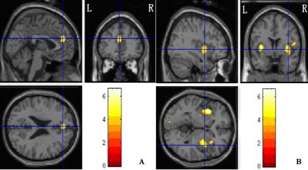
Figure 2.
Regional differences between PTSD group and control group. A showed that left Medial Frontal Gyrus (Brodmann's area 9) with significantly reduced gray-matter densities in PTSD group compared with controls. B showed that bilateral insulars (Brodmann's area 13) with significantly reduced gray-matter densities in PTSD compared with controls. Images were rendered onto orthogonal slices of the normal template magnetic resonance images.

