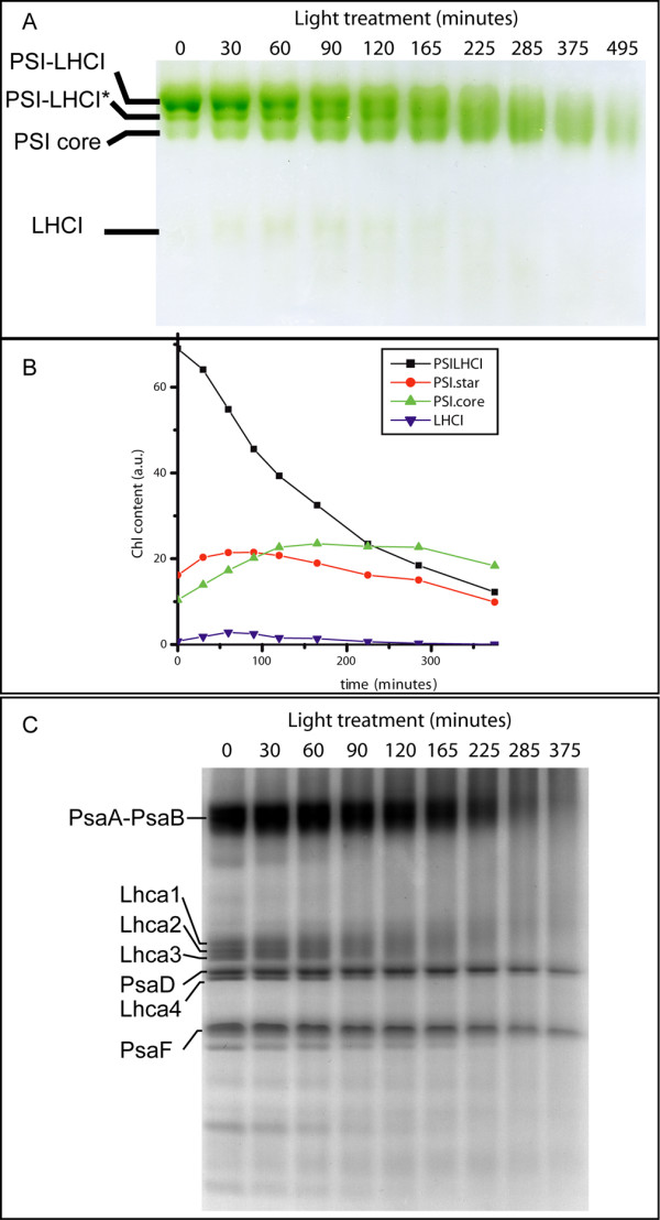
Figure 4.

LHCI is the first target in high light in PSI. PSI was treated with 1000 μE at 4°C and biochemically characterized during the treatment. The composition in different pigment protein complexes was analyzed by non denaturing Deriphat PAGE (A). (B) Densitometric quantification of different bands, normalized to the total Chl content of the sample before treatment. C) Polypeptide composition of PSI particles during the illumination was analyzed by SDS PAGE. The bands corresponding to Lhca, PsaA-B, PsaD and PsaF polypeptides, as identified by western blotting analysis, are evidenced. Other bands corresponding to PSI core polypeptides are indicated.
