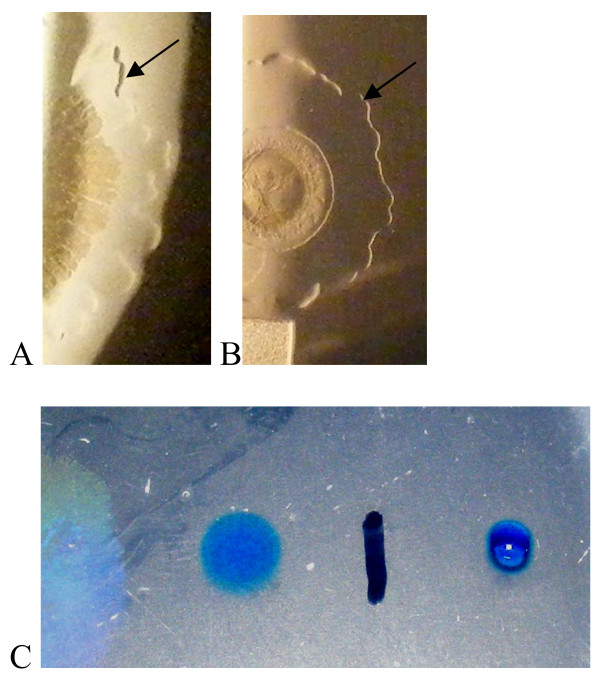
Figure 4.
A wetting agent is present beyond the edge of the swarm. Colony photography using reflected light (A, B) illustrating the presence of a wetting agent (arrows) preceding the spreading colony on (A) FW medium with succinate and NH4Cl as C, N source. B) Colony spread is limited by 500 μg/L CR, but wetting agent spreads as above. C) Drop collapse assay using dilute methylene blue solution showing the reduced surface tension in the wetting agent zone (left of the black line).

