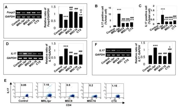
Figure 4.
The numbers of Foxp3+ cells and Th17 cells contributed to pathological process in MRL/lpr mice (A) Semi-quantitative RT-PCR confirmed decreased Foxp3 gene expression in bone marrow of MRL/lpr mice and increased Foxp3 expression in the treatment groups. The results were representative of five independent experiments ([[[P<0.001 vs. control; [[P<0.01 vs. control; ###P<0.001 vs. MRL/lpr; #P<0.05 vs. MRL/lpr; $$P<0.01 vs. MSCT). (B) Immunohistochemical staining with anti-IL17 antibody indicated that number of IL17 positive cells (mean±SD, arrows) was significantly increased in bone marrow (BM) of MRL/lpr mice (n=6). MSCT (MSC9, n=6; MSC16, n=6), as well as CTX treatment (n=6), significantly reduced IL17-positive cells in MRL/lpr bone marrow, but still showed higher level than that in control group ([[[P<0.001 vs. Control, ###P<0.001 vs. MRL/lpr). (C) Immunohistochemical staining using anti-IL17 antibody showed that number of IL17 positive cells (mean±SD, arrows) was significantly increased in spleen of MRL/lpr (n=6) compare to control group (n=6) and treatment group (MSC9; n=6, MSC16; n=6, CTX; n=6) ([[[P<0.001 vs. Control; ###P<0.001 vs. MRL/lpr). (D) Semi-quantitative RT-PCR revealed high expression of IL17 in bone marrow of MRL/lpr and this increased level of IL17 was decreased in MSCT and CTX treatment groups. The results were representative of five independent experiments ([[[P<0.001 vs. control; ###P<0.001 vs. MRL/lpr). (E) Flow cytometry revealed that MRL/lpr mice had significantly increased level of CD4+IL17+ T lymphocytes in spleen compared to control group. The CD4+IL17+ cells were markedly decreased in MSCT and CTX groups. (F) Semi-quantitative RT-PCR confirmed increased IL17 expression in spleen of MRL/lpr and reduced IL17 expression in the treatment groups. The results were representative of five independent experiments ([[[P<0.001 vs. control; ###P<0.001 vs. MRL/lpr; $P<0.05 vs. MSCT).

