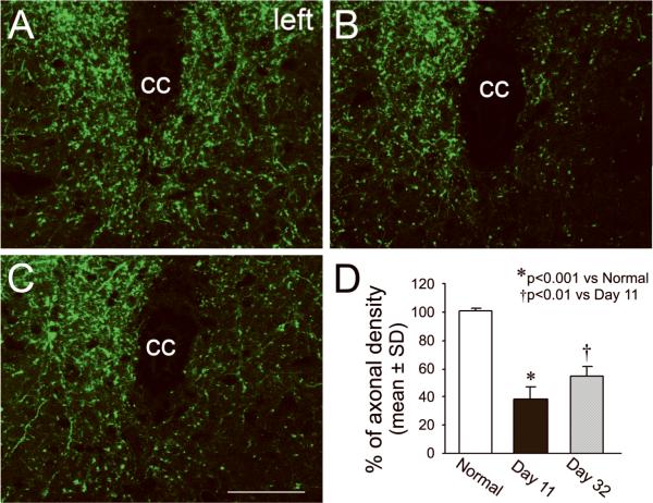
Figure 2. Single layer confocal images of CST axons in the gray matter of the cervical cord.
In normal CST-YFP mice, the CST axons are visible in the spinal gray matter on vibratome traverse sections with fluorescent microscopy (A). In mice with right MCAo, damaged axons were degenerated in the left side of spinal cord 11 days after stroke (B and D, p<0.001). However, axonal density significantly recovered 32 days after stroke (C and D, p<0.01). cc: central canal. Scale bar=50 μm.

