Figure 3.
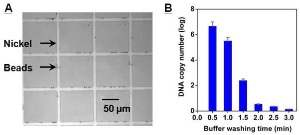
MMS device performance. A) Optical micrograph of the trapped magnetic beads. The beads are preferentially captured at the edge of the nickel patterns, where the magnetic field gradients are the highest. B) Real-time PCR measurement of eluted DNA as a function of wash duration. After 3 minutes of washing at 20 mL/hr in the microchannel, all weakly- and unbound DNA were eluted from the MMS device. The measurements were taken in triplicate and real-time PCR was performed in 20 μL volume with forward and reverse primers at 500 nM.

