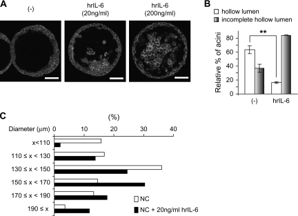
Fig. 5.
Effects of exogenous IL-6 on morphology of control RWPE-1 cells in 3D culture. (A) RWPE-1/NC cells were cultured in 3D with or without the indicated amounts of human recombinant (hr) IL-6 for 9 days, fixed, stained with 4′,6-diamidino-2-phenylindole (blue) to detect nuclei and rhodamine–phalloidin (red) to detect filamentous actin. Cells were visualized using confocal fluorescence microscopy. Bar, 50 μm. (B and C) Hollow lumen formation and acinar diameters of RWPE-1/NC cells cultured in 3D in the absence or presence of 20 ng/ml IL-6 were determined as in Figure 2. The data are presented as mean percentage ± SD of total structures counted. Student's t-test, **P < 0.01. The color version of this figure is available at Carcinogenesis Online.

