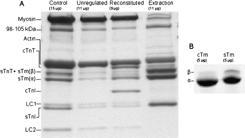
Fig. 1.
a Myofibril reconstitution preparation where the endogenous Tm and Tn were removed and replaced with sTm and cTn. Samples retained at each stage of the reconstitution protocol were run on a 12% Tris-HCl SDS gel and stained with coomassie blue. From left, the lanes shown are: control myofibrils, myofibrils after regulatory proteins have been extracted, myofibrils following reconstitution with sTm and cTn, the supernatant from the extraction stage of the protocol. The approximate amount of protein loaded into each lane is indicated. The TnC subunits were not visible due to poor stain retention of the protein at the concentrations present on the gel. The band present at 98–105 kDa is most likely α-actinin [38]. b Comparison of isoform content of the cTm and sTm used as replacement proteins. Samples (5 μg) were run on a 4–20% Tris-HCl SDS gel. The cTm is composed of 99% α-Tm, and sTm is 85% α-Tm and 15% β-Tm

