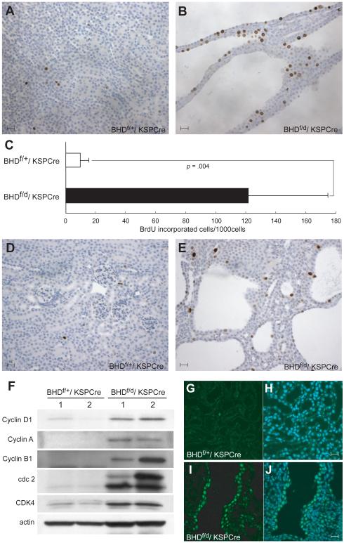
Fig. 4. Evidence of hyperproliferating cells in the kidney of BHDf/d/ KSP-Cre mice.
5′-Bromo-2′-deoxyouridine (BrdU) (100 μg/g body wt) was injected intraperitoneally at P14 into BHDf/+/KSP-Cre and BHDf/d/KSP-Cre mice 2 hr before euthanization. A) BrdU staining in BHDf/+/KSP-Cre mouse kidney. B) BrdU staining in BHDf/d/KSP-Cre mouse kidney. C) Greater than 10-fold more BrdU incorporation was seen in BHDf/d/KSP-Cre mouse kidneys compared with BHDf/+/KSP-Cre mouse kidneys (n=5 for each group, mean=121.8 per 1000 cells versus 9.6 per 1000 cells, difference=112.2, 95% CI=59.3 to 165.0; Welch’s t test:P=.004). 1000 cells per field were counted in five randomly selected fields from two mice for each group. Data are represented as means and 95% confidence intervals. D) Phospho-histone H3 staining detects few G2/M phase cells in 3-week-old BHDf/+/KSP-Cre kidney. E) Phospho-histone H3 staining in 3-week-old BHDf/d/KSP-Cre mouse kidney shows more G2/M phase cells. F) Immunoblotting analysis of proteins extracted from kidneys of 3-week-old BHDf/+/KSP-Cre and BHDf/d/KSP-Cre mice. Cell cycle promoting proteins are highly expressed in BHDf/d/KSP-Cre mouse kidneys. Data represent typical results for two mice of each genotype from at least three independent experiments. G) Cyclin D1 staining (green) in the kidney of a 2-week-old BHDf/+/ KSP-Cre mouse is weak. H) Merged image of Cyclin D1 with 4′-6-Diamidino-2-phenylindole (DAPI) nuclear staining (blue). I) Nuclear Cyclin D1 staining (green) is seen in cells lining the dilated tubules in the kidney of a 2-week-old BHDf/d/KSP-Cre mouse. J) Merged image of Cyclin D1 with DAPI nuclear (blue) staining. Scale bar=20 μm.

