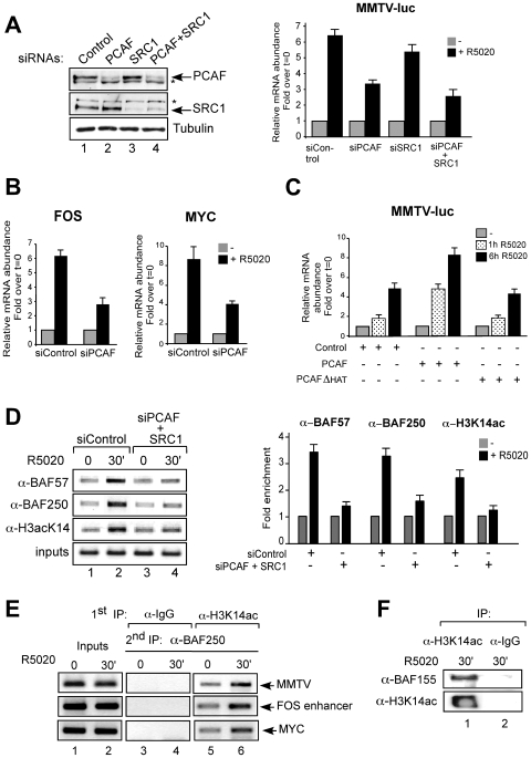
Figure 3. Acetylation on histone H3K14 by PCAF is essential for hormonal transactivation in T47D-MTVL cells.
(A) Left: Cells were transfected with the indicated siRNAs and the levels of PCAF, SRC1, and tubulin were determined by Western blotting. The asterisks indicate inespecific bands. Right: Cells were transfected with the indicated siRNAs and treated with 10 nM R5020 for 6 h. RNA was extracted, cDNA was generated and used as template for real time PCR with luciferase oligonucleotides. The values represent the mean and standard deviation from 3 experiments performed in duplicate. (B) Cells were transfected with siRNAs, treated with 10 nM R5020 as indicated and the levels of c-Fos and c-Myc mRNAs were analyzed by RT–PCR. The values represent the mean and standard deviation from 2 experiments performed in duplicate. (C) Cells were transfected with PCAF or PCAF HAT mutant (PCAFΔHAT), treated with hormone as indicated and transcription from the MMTV promoter was determined as in (B). An empty vector was used as control. The values represent the mean and standard deviation from 2 experiments performed in duplicate. (D) Left: Cells transfected with the siRNAs and treated with hormone as indicated were subjected to ChIP assays with α-BAF57, α-BAF250 and α-K14acH3. The precipitated DNA fragments were subjected to PCR analysis to test for the presence of sequences corresponding to the MMTV nucleosome B. A representative of three independent experiments is shown. Right: quantification of the results by real time PCR from two experiments performed induplicate. (E) Cells were treated with R5020 as indicated and subjected to re-ChIP assays with α-H3K14ac and IgG (first IP) followed by α-BAF250 for the second IP. Precipiated DNA was analysed by PCR for the presence of sequences corresponding to the MMTV, c-Fos and c-Myc PR binding regions. (F) Cells were treated for 30 min with R5020, lysed and chromatin was immunoprecipitated either with α-H3K14ac or with normal rabbit IgG as a negative control (IgG). IPs were analyzed by western blot using α-BAF155 and α-H3K14ac.

