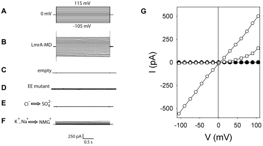
Figure 2. Electrophysiological analyses of ion transport in proteoliposomes by ‘dip-tip’ method.
A, Voltage step protocol from a holding potential of 0 mV to various voltages (ranging from −105 mV to +115 mV), back to 0 mV. B, C, D, Current traces for LmrA-MD (B) or empty liposomes without protein (C) or EE LmrA-MD (D) in the presence of 10 mM NaCl. E, F, Current traces for LmrA-MD in the presence of SO4 2− instead of Cl− (E), or NMG+ instead of Na+ and K+ (F). G, I–V curves from the traces in (B–F) (○, LmrA-MD; •, replacement of Cl− by SO4 2−; □, replacement of K+/Na+ by NMG+; ▿, EE; ▵, empty liposomes; the latter two traces are hidden behind •). (n = 15)

