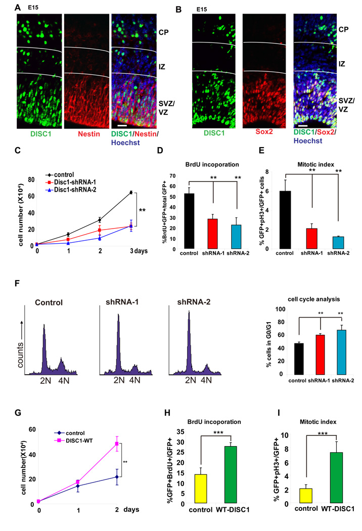
Figure 1. DISC1 regulates progenitor cell proliferation in vitro.
(A) E15 embryonic brain sections were co-stained with anti-DISC1 and anti-Nestin antibody. Scale bar=20 µm.
(B) E15 embryonic brain sections were co-stained with anti-DISC1 and anti-Sox2 antibody. Scale bar=20 µm.
(C) Cell proliferation is reduced in AHP cells infected with lentivirus expressing DISC1 shRNAs. GFP expression is used as a marker for viral infection. Both DISC1 shRNAs significantly reduced cell proliferation (n=3, p<0.01).
(D) BrdU incorporation is decreased in DISC1 knockdown cells. AHPs infected with control, DISC1 shRNA-1, or shRNA-2 lentivirus were pulse labeled with 10 µM BrdU and stained with BrdU antibody. The percentage of GFP positive cells that are also BrdU positive cells is shown (n=4, p<0.01).
(E) Mitotic index is reduced in DISC1-silenced AHPs. The percentage of GFP positive cells that are also pH3 positive is shown (n=3, p<0.01).
(F) Histograms for FACS analysis of N2a cells transfected with DISC1 shRNAs. Bar graph depicts the percentage of GFP positive cells in G0/G1 (n=3, p<0.01).
(G) Cell proliferation is increased in DISC1 overexpressing cells. AHPs were infected with either control or DISC1-WT lentivirus. Cell number was counted for 2 days (n=3, p<0.01).
(H) BrdU incorporation is increased in DISC1-overexpressing AHPs. The percentage of GFP positive cells that are also BrdU positive cells is shown (n=4, p<0.001).
(I) Increased mitotic index in DISC1 overexpressing AHPs. The percentage of GFP positive cells that are also pH3 positive is shown (n=4, p<0.001).

