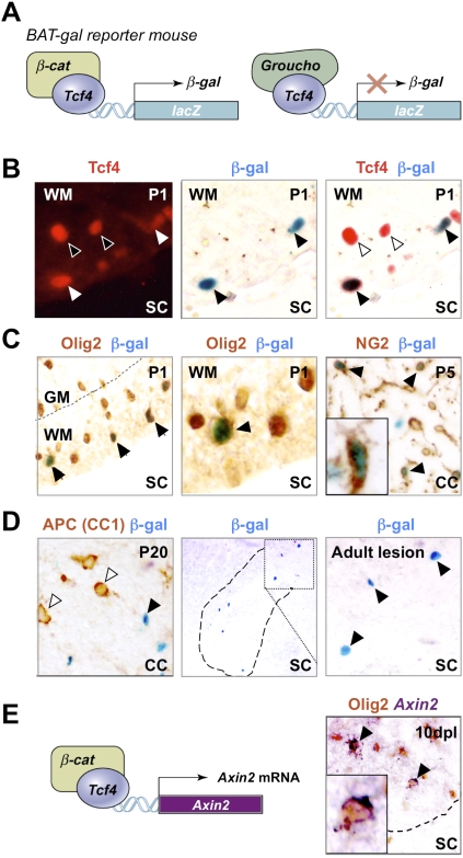
Figure 4.
Use of BAT-gal mice demonstrates that Tcf4 functions in catenin-dependent Wnt signaling during brain and spinal cord developmental myelination and remyelination of adult spinal cord. (A) Schematic for Tcf4–β-catenin-driven β-galactosidase (β-gal) reporter expression in BAT-gal mice. (C) β-gal is expressed in a subset of Olig2 cells in BAT-gal reporter mice in presumptive white matter only of P1 SC, and in a subset of NG2 cells in P5 CC, suggesting that the Wnt pathway is active in these cells. β-gal is expressed in a subset of Tcf4 cells in BAT-gal Wnt reporter mice P1 SC white matter (B), but is not expressed in cells expressing the Wnt pathway inhibitor APC (CC1) in P20 CC (D). (D) β-gal is re-expressed in a demyelinated lesion at 5 dpl following injection of lysolecithin into BAT-gal spinal cord. Filled arrowheads refer to BAT-gal-positive cells. Unfilled arrowheads show Tcf4 or CC1 staining cells that are BAT-gal-negative. (E) Schematic for Tcf4–β-catenin-driven Axin-2 mRNA expression. Axin2 is a known target of Wnt signaling (Lustig et al. 2002) that is globally up-regulated by activation of the Wnt pathway. Endogenous Axin-2 mRNA is expressed in a subset of Olig2 cells at 10 dpl in a remyelinating spinal cord lesion.

