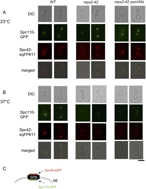
Figure 8.
SPBs are inserted into the nuclear envelope in mps2-42 pom34Δ cells. (A,B) Analysis of wild-type, mps2-42, and mps2-42 pom34Δ cells with SPC110-GFP SPC42-eqFP611 by fluorescence and phase contrast (DIC) microscopy at 23°C (A) and 37°C (B) (2 h). Note that in B Spc110-GFP is only associated with one of the two Spc42-eqFP611-marked SPBs of mps2-42 cells. This is the typical phenotype of cells with a defect in duplication plaque insertion (Schramm et al. 2000; Jaspersen and Winey 2004). Bars, 5 μm. (C) Shown is a cartoon of the SPB with the localization of Spc42 and Spc110 relative to the nuclear envelope (NE) (Adams and Kilmartin 1999).

