Figure 3.
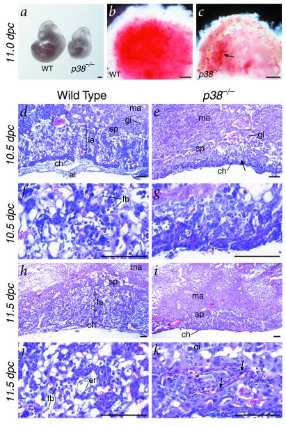
Morphology and histology of wild-type and p38α mutant embryos and placentas. (a) Gross morphology of a wild-type and p38α mutant littermate at 11.0 dpc, showing developmental retardation of the homozygote. (b and c) Ventral view of the placentas of a wild-type and p38α mutant, showing apparent decreased vascularization of the mutant placenta, as shown by its paler red color (arrow in c). (d–k) Hematoxylin and eosin-stained sections of placentas from wild-type (d, f, h, and j) and p38α homozygous mutant littermates (e, g, i, and k). (d and e) At 10.5 dpc, the developing labyrinth and spongiotrophoblast layers found in the wild type (d) are significantly decreased in thickness in the p38α mutant (e, arrow). (f and g) High-power views show a nearly complete lack of circulating fetal blood cells in the mutant. (h and i) By 11.5 dpc, the labyrinth layer is essentially missing and the spongiotrophoblast layer is greatly diminished in the p38α mutant compared with wild-type. (j and k) High-power views show that the placental vasculature with differentiated endothelial cells and circulating fetal blood cells (j) is nearly completely abolished in the p38α mutant, which instead contains apparent necrotic regions (k, arrows). (Scale bars in a–c represent 0.5 mm; bars in d–k represent 100 μm.) Abbreviations: al, allantois; ch, chorionic plate; en, endothelial cells; fb, fetal blood cells; gi, trophoblast giant cells; la, labyrinthine trophoblast; ma, maternal decidual tissue; sp, spongiotrophoblast; WT, wild type.

