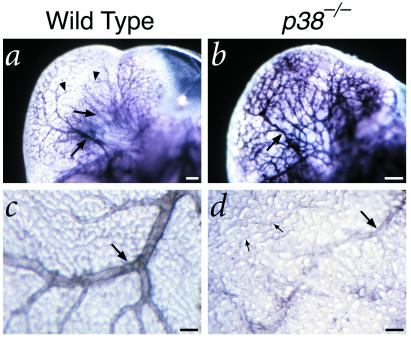
Figure 5.
PECAM whole-mount immunohistochemistry of embryos and visceral yolk sacs at 10.5 dpc. (a and b) Vasculature of the developing brain in a wild-type littermate (a) and p38α mutant embryo (b). Note that the tree-like architecture of the major vessels (arrows) connecting to minor branches (arrowheads) in the wild-type differs significantly from the relatively uniform size and disorganized pattern of the vasculature in the mutant embryo. (c and d) Vasculature of the visceral yolk sac in a wild-type littermate (c) and p38α mutant embryo (d). Although the wild-type yolk sac displays major vessels (arrows) and branches, the mutant yolk sac has relatively few major vessels with abnormal morphology (arrow), and retains a primitive capillary plexus (small arrows).

