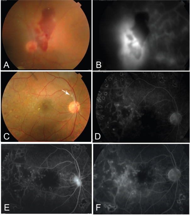
Figure 3.
A 40 years old patient with PDR and vitreous hemorrhage (A). B, The fluorescein angiogram showed evident neovascularization at the disc (NVD) with evident nasal ischemia. C, One month following bevacizumab injection there was near complete absorption of vitreous hemorrhage with a fibrous tuft arising from the optic disk (arrow). The macula looks dry with marks of previous grid. The angiogram (D) showed near complete regression of the neovascularization. E, three months following single injection, the angiogram showed evidence of reperfusion of NVD. F, 2 weeks following re-injection, the angiogram showed again complete regression of the NVD.

