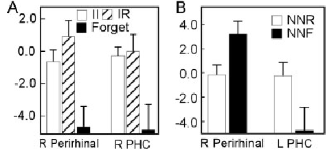
Figure 2.

Predictive memory effects at time of study (A) and at time of test (B). At study, II (white) and IR (hashed) trials were combined and their activity was contrasted with the activity associated with subsequently forgotten trials (black). Two MTL regions, one within right perirhinal cortex and one within right parahippocampal cortex, exhibited an overall predictive memory effect. At test, activity associated with new items remembered outside the scanner (NNR; white) was contrasted with activity associated with new items forgotten outside the scanner (NNR; black). Two MTL regions, one within right perirhinal cortex and one within left parahippocampal cortex showed predictive memory effects. The vertical axis represents activity (sum of beta coefficients) and error bars indicate the standard error of the mean.
