Figure 1.
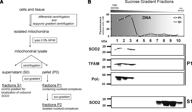
Isolation of human nucleoids from Jurkat cells. A) Tissue or cells were disrupted with a Dounce homogenizer in isoosmotic mitochondrial isolation buffer. Mitochondria were isolated with differential centrifugation followed by an isopycnic gradient centrifugation step as described in methods. Pure mitochondria were lysed with the nonionic detergent Nonidet P-40 and fractionated into supernatant (S0) and pellet (P0) by centrifugation. Pure nucleoids were isolated from the pellet by 2 sequential sucrose step gradients (P1 and P2; 75–20% sucrose). B) Nucleoids were purified from Jurkat mitochondria by a step gradient, and the P1 fractions were analyzed for their DNA (SYBR Green fluorescence)/protein distribution (Western blot analysis). MtDNA was concentrated in P1 fractions 1–3 (top panel). To identify nucleoid-containing fractions, Western blotting against TFAM and Polγ was performed. Nucleoid markers and SOD2 were found in the mtDNA-containing fractions (middle panel). Some unbound SOD2 was found in the low-density S fractions (bottom panel). Data shown are representative of 8 independent experiments.

