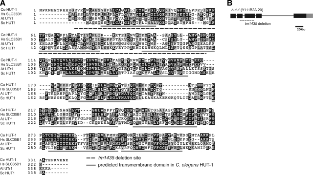
Figure 1.
HUT-1 protein and hut-1 gene. A) Alignment of SLC35B1 of C. elegans (Ce), Homo sapiens (Hs), Arabidopsis thaliana (At), and Saccharomyces cerevisiae (Sc). Identical amino acids are shaded in black; similar amino acids are shaded in gray. Transmembrane segments as predicted by the Phobius web server are underlined in gray. The deletion region in tm1435 is shown by a dashed underline. B) Structure of the hut-1 gene. Exons are indicated by boxes. Black and gray boxes are translated and untranslated regions, respectively. Dashed line indicates the deletion in tm1435.

