
Fig. 3. Expression of odontoblastic and ameloblastic differentiation markers in Tgfbr2fl/fl;Wnt1-Cre tooth germ cultured under a kidney capsule.
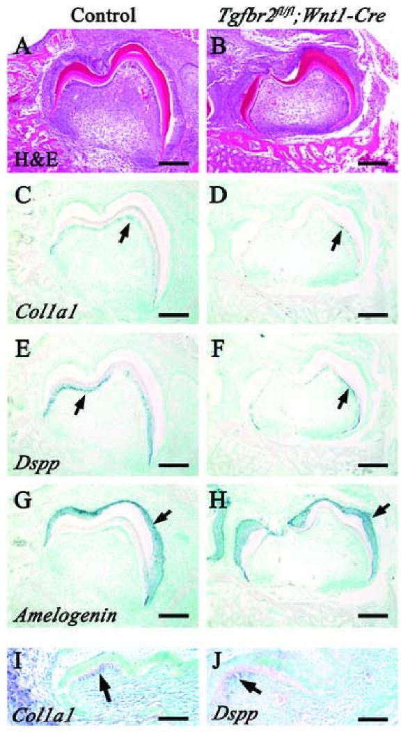
H&E staining and in situ hybridization of Dspp, Col1a1 and Amelogenin in wild type (A,C,E,G) and Tgfbr2fl/fl;Wnt1-Cre (B,D,F,H) samples after 14 days cultivation under a kidney capsule. (C,D) Expression of Col1a1 (arrows) in Tgfbr2fl/fl;Wnt1-Cre transplants is detectable in a similar pattern to wild type, although the level of the expression is reduced. (E,F) Expression of Dspp (arrows) is detectable in wild type and Tgfbr2fl/fl;Wnt1-Cre odontoblasts, but the level of expression is reduced in the Tgfbr2fl/fl;Wnt1-Cre sample. (G,H) The level and pattern of Amelogenin expression (arrows) in the Tgfbr2fl/fl;Wnt1-Cre sample are indistinguishable from wild type. (I,J) In situ hybridization of Col1a1 (I) and Dspp (J) in wild type samples after 5 days cultivation under a kidney capsule. Both Col1a1 and Dspp are already expressed in odontoblasts at the tip of the cusp at this stage (arrow). Scale bars: 100 μm in A-H; 50 μm in I and J.