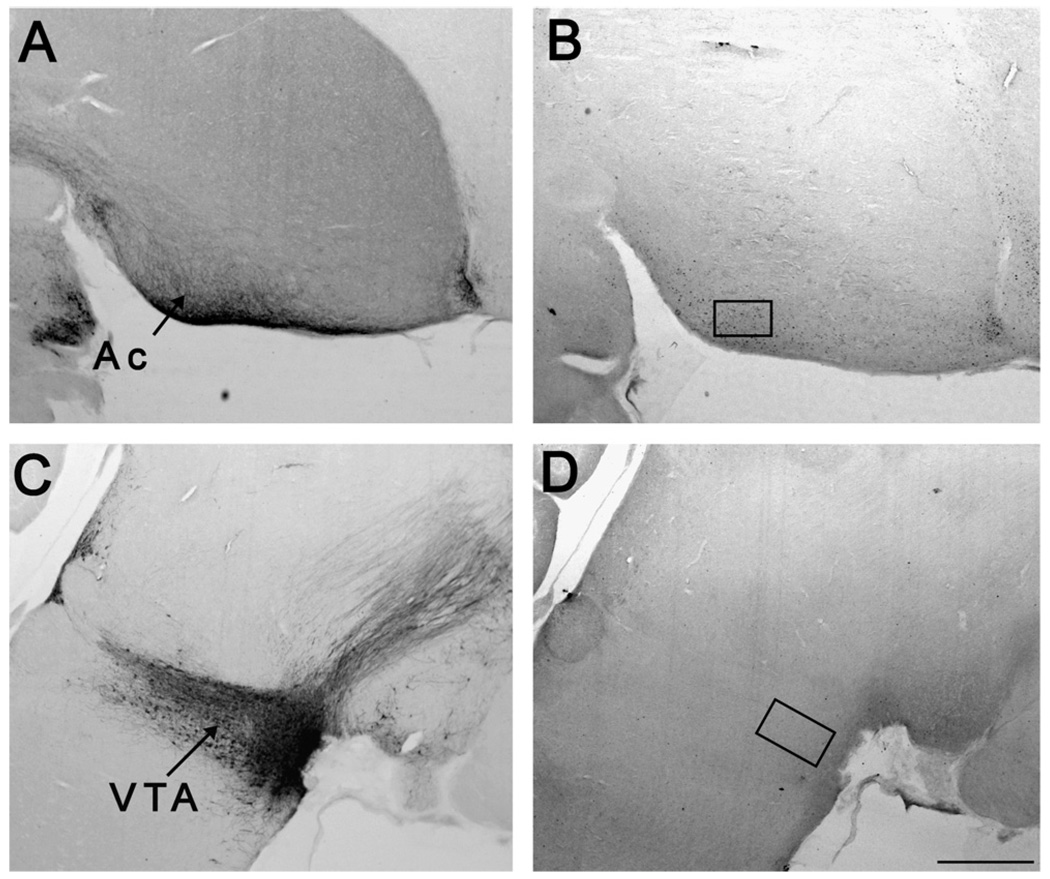
Fig. 2.
Placement of sampling regions within the reward system. Panels A and C contain sections subjected to tyrosine hydroxylase immunohistochemistry sliced in the sagittal plane. Panels B and D contain ZENK labeling. Panel B displays the location of the box for the nucleus accumbens (Ac; 190 µm × 305 µm), and panel D displays the ventral tegmental area (VTA; 210 µm × 368 µm). The more rostral portion of each photo is towards the right edge. Scale bar = 300 µm.

