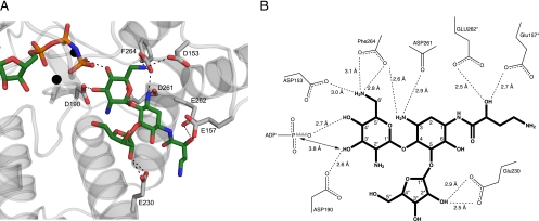
FIG. 2.
(A) Cartoon representation of the aminoglycoside-binding site of the butirosin A ternary complex of APH(3′)-IIIa. Residues interacting with butirosin A are drawn as gray sticks, and their hydrogen bond interactions with butirosin A are shown as dashed lines. The color scheme is the same as that shown in Fig. 1. (B) Schematic representation of the hydrogen bonding interactions between butirosin A and APH(3′)-IIIa. The distance between the γ-phosphate of the nucleotide and the reactive 3′-hydroxyl of the aminoglycoside substrate is also indicated. The asterisks denote the amino acid residues that underwent a conformation change in the amikacin-bound model of APH(3′)-IIIa.

