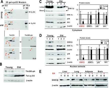
FIG. 2.
Hyperphosphorylation of cyclin D3 at Thr283 in livers of young mice correlates with high levels of GSK3β. (A) Hyperphosphorylated isoforms of cyclin D3 are observed only in livers of young mice. Nuclear extracts from livers of young and old mice were separated by 2D gel electrophoresis and probed with antibodies to cyclin D3. Positions of differentially expressed isoforms of cyclin D3 (a, b, and c) are shown by red arrows. For the bottom part, the membrane was reprobed with antibodies to the Thr283 phosphorylated isoform of cyclin D3. (B) The Thr283-ph isoform of cyclin D3 is abundant in young livers. Western blotting was performed with nuclear extracts from young and old mouse livers with antibodies to the Thr283-ph isoform of cyclin D3. The filter was reprobed with antibodies to total cyclin D3 and with antibodies to β-actin. (C and D) Protein levels of GSK3β are reduced in livers of old mice. Cytoplasmic (C) and nuclear (D) extracts from livers of young and old mice were examined by Western blotting with antibodies to GSK3β, GSK3α, PP1, p38, and β-actin. Short (30-s) and long (2-min) exposures of the cyclin D3 membrane are shown. The protein levels of the examined proteins were normalized to signals of β-actin. The bar graphs present a summary of three or four independent experiments. Error bars indicate standard deviations. (E) Treatment of old mice with GH increases protein levels of GSK3β in liver, lung, brain, and adipose tissues. Protein extracts from liver, lung, adipose, and brain tissues of young (Y), old (O), and GH-treated old mice were examined by Western blotting with antibodies against GSK3β. The membranes were reprobed with antibodies to β-actin. Since signals of cyclin D3 are different in the examined tissues, the optimal exposures are shown for the Western blotting with each tissue.

