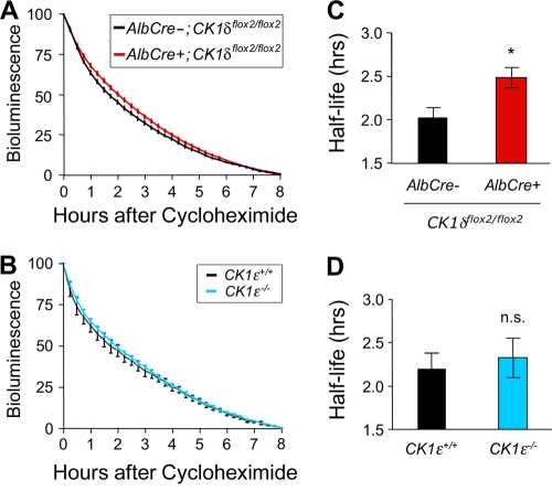
FIG. 10.
mPER2::LUC bioluminescence half-life is increased by disruption of CK1δ but is unaffected by the absence of CK1ɛ. (A and B) Liver explants were treated with cycloheximide (80 μg/ml) at the time of peak bioluminescence on the first day in vitro, and the decline in bioluminescence was monitored for 8 h. The data were normalized to the peak level of mPER2::LUC bioluminescence and the minimum level after 8 h of recording. Values are plotted as means ± the SEM. (A) Average bioluminescence profiles from explants from CK1δ-deficient liver (AlbCre+; CK1δflox2/flox2; mPer2Luc/+) (n = 12) and control liver (AlbCre−; CK1δflox2/flox2; mPer2Luc/+) (n = 7) tissues. (B) Average bioluminescence profiles from liver explants from CK1ɛ-deficient livers and wild-type (CK1ɛ+/+; mPer2Luc/+) controls (n = 8 per genotype). (C) The calculated bioluminescence half-life was significantly (*, P < 0.025) increased in CK1δ-deficient livers, relative to controls lacking Cre recombinase. (D) mPER2::LUC bioluminescence half-life was unaltered in CK1ɛ-deficient livers.

