FIG. 3.
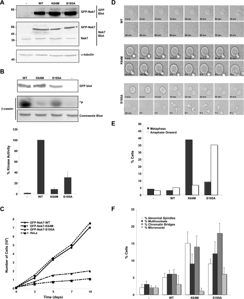
Cells stably expressing kinase-inactive Nek7 exhibit defective cell cycle progression. (A) Cell lysates were prepared from parental HeLa cells (−) or HeLa cells stably expressing GFP-Nek7 constructs, as indicated, and were subjected to SDS-PAGE and Western blot analysis with antibodies against GFP, Nek7, and α-tubulin. Molecular masses (kDa) are indicated on the left. WT, wild type. (B) Cell lysates were subjected to immunoprecipitation with anti-GFP antibodies, as described in the legend for Fig. 1A. The amount of kinase precipitated was determined by Western blotting with anti-GFP antibodies and the immunoprecipitates used for kinase assays, with β-casein as a substrate. Products were analyzed by SDS-PAGE, Coomassie blue staining, and autoradiography (32P). Activity levels of the different Nek7 constructs are expressed as a percentage of the wild-type activity normalized to the amount of precipitated protein. The data are shown as means (±standard deviations) from three separate experiments. (C) Growth curves of HeLa cells stably expressing GFP-Nek7 constructs are shown; data represent the means from three separate experiments. (D) Mitotic HeLa cells stably expressing GFP-Nek7 constructs, as indicated, were analyzed by time-lapse bright-field microscopy. Images are presented from metaphase onwards, with time indicated on each panel. (E) The outcome of each live cell imaged as in panel D was scored according to whether they were delayed in metaphase or late mitosis prior to undergoing apoptosis or whether they progressed normally. A total of 40 to 50 cells were scored for each cell line. (F) Parental HeLa cells (−) or HeLa cells stably expressing GFP-Nek7 proteins, as indicated, were fixed and stained with α-tubulin antibodies to detect the microtubule network and Hoechst 33258 to detect DNA. The percentage of cells with abnormal mitotic spindles or cells that were multinucleated, micronucleated, or still attached by chromatin bridges was scored. Data represents means (±standard deviations) from counts of at least 150 cells in three independent experiments.

