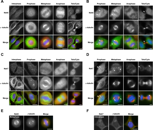
FIG. 6.
In mitosis, Nek6 localizes to spindle microtubules and spindle poles, while Nek7 localizes only with spindle poles. (A) HeLa cells were processed for immunofluorescence microscopy with Nek6 (red in merge) and α-tubulin (green in merge) antibodies. Telo/Cyto, telophase/cytokinesis. (B) HeLa cells were treated with extraction buffer for 30 s before being fixed and permeabilized in methanol and processed for immunofluorescence microscopy, as described in the legend for panel A. Arrowheads denote spindle pole or midbody staining. (C and D) Same process as described in legends for panels A and B, respectively, except using Nek7 (red) antibodies. (E and F) HeLa cells were fixed and processed for immunofluorescence microscopy with Nek6 or Nek7 (red) antibodies, as indicated, and γ-tubulin (green) antibodies. In all panels, DNA was stained with Hoechst 33258 (blue), and merged images are shown. Scale bars, 10 μm.

