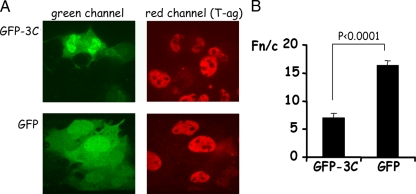
FIG. 4.
Overexpression of GFP-3C can reduce nuclear accumulation of T-ag in COS-7 cells. COS-7 cells grown on coverslips were transfected to express either GFP alone or GFP-3C and fixed 18 h later with 4% formaldehyde followed by permeabilization of membranes with 0.2% Triton X-100. Cells were probed with rabbit polyclonal antibody to simian virus 40 T-ag (Santa Cruz Biotechnology) followed by detection with Alexa Fluor 568-conjugated antibodies to rabbit immunoglobulin. Coverslips were mounted on glass slides, and CLSM and image analysis were performed as described in the legend to Fig. 1. (A) Single images of cells expressing GFP alone or GFP-3C as indicated. The green channel (GFP and GFP-3C) is on the left, and the red channel (T-ag) is on the right. (B) Images such as those shown in panel A were analyzed as described in the legend to Fig. 1. Results are means ± standard errors of the mean, where n represents ≥15.

