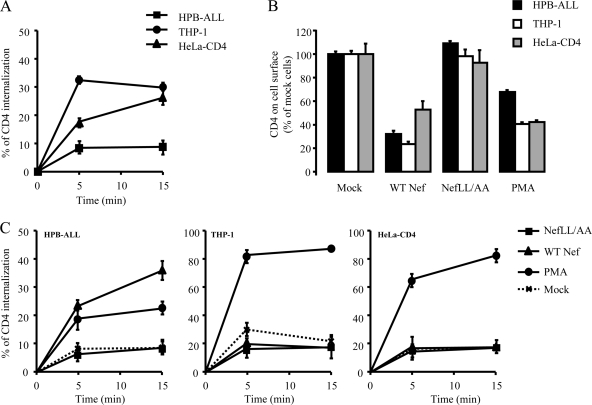
FIG. 1.
Effect of Nef and PMA on CD4 internalization in lymphoid, myeloid, and HeLa-CD4 cells. Cell surface expression of CD4 at steady state (B) as well as the CD4 internalization rate (A, C) were analyzed by FACS in lymphoid (HPB-ALL), myeloid (THP-1), and HeLa-CD4 cells expressing WT or mutant Nef-GFP or treated with PMA. (A) Cells were stained at 4°C with an anti-CD4 antibody and incubated at 37°C for 0, 5, and 15 min to allow for CD4 internalization. Cells were then cooled down and stained with Cy5-coupled secondary antibody. The percentage of internalized CD4 at each time point was calculated as described in Materials and Methods. Transfected cells were gated on the basis of GFP expression. (B) Steady-state levels of cell surface CD4 in cells expressing Nef-GFP (gated on the basis of GFP expression) or treated with PMA. Results are expressed as the percentage of cell surface CD4 on mock-treated/transfected cells. (C) CD4 internalization was assessed, as described in panel A, on transfected or PMA-treated cells. Values are the means of three independent experiments; error bars represent 1 standard deviation from the mean.

