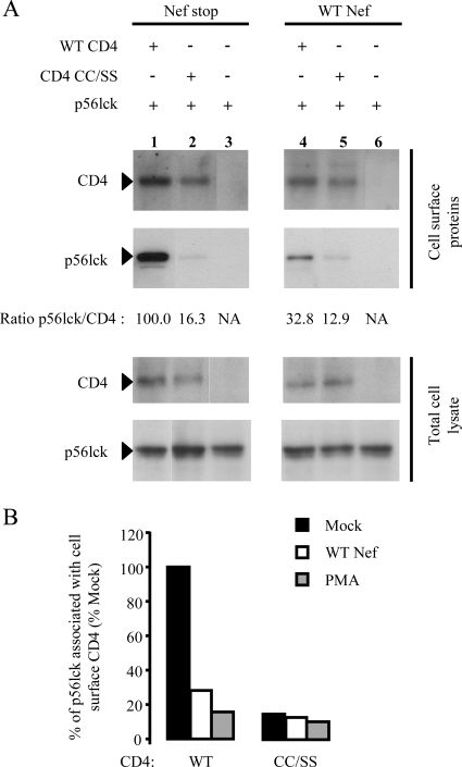
FIG. 6.
Nef-induced decrease of CD4/p56lck association at the plasma membrane. (A) HeLa cells coexpressing p56lck and either WT CD4 or CD4 CC/SS were transfected to achieve Nef expression or treated with PMA. After biotinylation of the cell surface proteins, biotinylated CD4 was immunoprecipitated, as described in Materials and Methods, and both CD4 and p56lck were visualized by Western blotting, followed by immunodetection in the precipitated material (top) or in cleared lysates (20 μg of protein/lane; bottom). The intensity of the CD4 and p56lck bands was quantified by densitometry using NIH Image software, and the p56lck signal was normalized to that of cell surface CD4 (NA, not applicable). (B) Plotted data represent the percentage of the CD4-associated p56lck signal intensity relative to that of mock-transfected or mock-treated cells expressing WT CD4 (100%) and are representative of three independent experiments.

