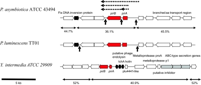
FIG. 1.
Comparison of the genomic organization of the pirAB operons in two species of Photorhabdus and one strain of Yersinia. PirAB genes are filled in red. Note that the presence of mobile element-related sequences (filled in black) and the G+C content of the regions suggest recent horizontal acquisition of these genes. Also, note that the Yersinia pirAB genes are tightly linked to a type I secreted metalloprotease operon, suggesting a larger pathogenicity island. ERIC sequences in the Photorhabdus operons are indicated by vertical arrows. Dotted horizontal arrows show the PCR-generated clones tested in this work.

