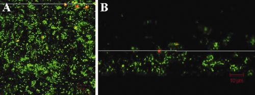
FIG. 1.
Top-down projection (A) and cross-sectional view (B) of a summer biofilm culture, with C. parvum oocysts attached at the biofilm surface. Biofilm cells are stained green with SYTO 9; oocysts are stained red with Cy3. The white line in panel A indicates the location of the cross section shown in panel B. Direction of water flow is from right to left. The biofilm is approximately 24-μm thick; oocysts are located 16 μm above the biofilm base.

