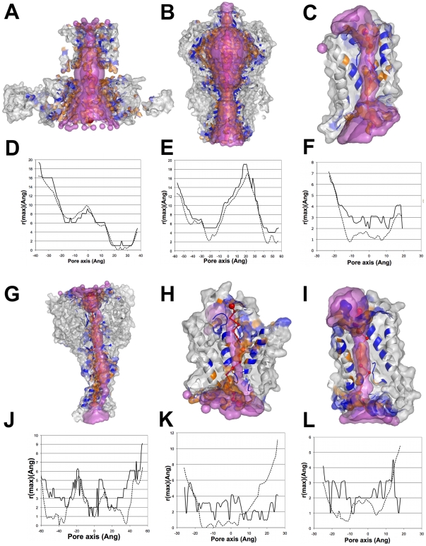
Figure 6. Comparisons of channel cavities identified by PoreWalker and HOLE and corresponding 1Å step profile diameters.
(A)&(D) MthK potassium channel (1lnq), (B)&(E) MscS voltage-modulated mechanosensitive channel (2oau); (C)&(F) bovine aquaporin-0 (1ymg), (G)&(J) ASIC1 acid-sensing ion channel (2qts); (H)&(K) Amt-1 ammonium channel (2b2f); (I)&(L) SoPiP2;1 water channel (1z98). PoreWalker cavities are shown as y-coordinate>0 only of the protein section along the xy-plane, where the x-axis corresponds to the pore axis. Pore-lining atoms and residues are coloured in orange and blue, respectively, and the rest of the protein is shown in light grey. Red spheres represent pore centres at 3Å steps and their size is proportional to the pore diameter at that point. HOLE cavities are shown as purple surface. PoreWalker and HOLE profile diameters are shown as solid and dotted lines, respectively. Corresponding R2 values are 0.958, 0.951, 0.814, 0.450, 0.017 and 0.000 for 1lnq, 2oau, 1ymg, 2qts, 2b2f and 1z98, respectively.

