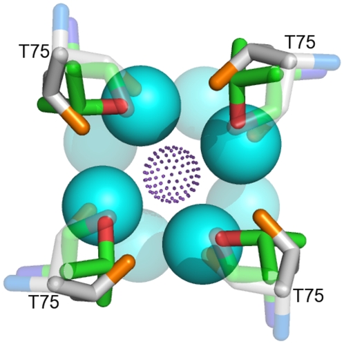
Figure 9. Superposition of the 3D-structures of Thr75s from the low-K+ (green/red/blue colour scheme) and high-K+ (white/orange/blue colour scheme) KcsA channels, top view.
Water molecules from the low-K+ structure are shown in light blue. Violet dots represent a K+ ion. Thr side chain atoms and the four water molecules interacting with them are shown as opaque. Thr backbone atoms and the remaining four water molecules of the K+ hydration shell are shown as transparent.

