Fig. 1.
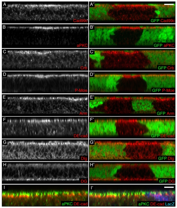
hpo loss of function induces an increase in apical polarity proteins in wing imaginal cells. (A-H′) Transverse sections of third instar wing discs containing clones of hpo mutant cells (marked by absence of GFP). Apical is to the top in this and all subsequent XZ sections. Scale bar: 10 μm. (A′-H′) Merged images of A-H with GFP images (green). (I,I′) Transverse section of a third instar wing disc containing clones of hpo5.1 mutant cells (marked by absence of β-gal) stained for aPKC (green) and DE-cad (red). (I′) Merged image of I with β-gal (blue). Scale bar: 5 μm. hpo mutant cells have an increased apical staining of the following apical determinants: Cad99c (A,A′), aPKC (B,B′), Crb (C,C′), P-Moe (D,D′), Arm (E,E′) and DE-cad (F,F′). By contrast, they do not show an increase of the septate junction protein Dlg (G,G′) or the basal marker Dystroglycan (DG; H,H′). Despite their respective increase, the proteins aPKC and DE-cad remain in non-overlapping membrane domains (I,I′).

