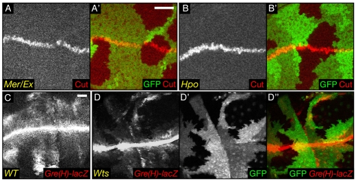
Fig. 5.
Downregulation of Notch activity in Hpo signalling-deficient cells. (A-D′) XY sections of wing imaginal discs. (A′,B′) Merged images of A and B with GFP (green). (D″) The merged images of D (red) and D′ (green). Scale bar: 20 μm. mer;ex (A,A′) and hpo (B,B′) mutant cells do not show an increase in Cut staining at the dorsoventral boundary. Compared with WT cells (C), wts mutant cells (D,D′) show a decrease in β-galactosidase staining monitoring the activity of the Notch reporter Gre(H)-lacZ in the pouch, and also more weakly at the dorsoventral boundary.

