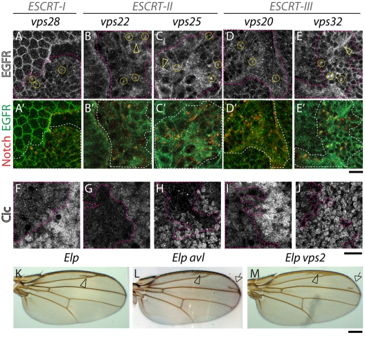
Fig. 4.
EGFR accumulation and EGFR signaling is overactivated in ESCRT mutants. (A-E′) ESCRT-I, ESCRT-II and ESCRT-III mosaic eye discs stained with anti-EGFR (A-E) and Notch (merged in A′-E′); Compared with WT tissue, in mutant tissue (delimited by pink dashed lines) EGFR accumulation is seen in intracellular puncta (examples are encircled in A-E) that are Notch positive. Note that compared with Notch, the majority of EGFR is still detected in other cellular areas (arrowheads in B-C,E). (F-J) ESCRT-I, ESCRT-II and ESCRT-III mosaic eye discs stained with an antibody to detect expression of Capicua (Cic), a nuclear negative regulator of EGFR signaling. Compared with WT tissue, Cic expression is reduced in ESCRT mutant cells (delimited by pink dashed lines), indicating higher levels of EGFR signaling activation. Adult wings carrying Ellipse mutations, an activated form of EGFR (Elp; K). Animals also heterozygous for avl (Elp avl; L) or vps2 (Elp vps2; M) show extra wing vein material, indicative of excessive EGFR signaling (arrowhead in K). The frequency of extra veins is increased in Elp avl wings (+82% n=60; arrows in L) and, to a lesser extent in Elp vps2 (+38% n=19; arrows in L-M), indicating that excess EGFR signaling is enhanced by reducing endocytic gene dosage. Scale bars: 10 μm (A-J), 100 μm (K-M).

