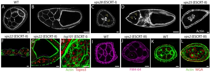
Fig. 5.
ESCRT complexes regulate actin cytoskeleton stability and membrane integrity. (A-E) Stage 6 (A,C,E) and stage 10 (B,D) egg chambers stained with phalloidin to reveal the cortical actin cytoskeleton. Compared with WT (A,B), egg chambers containing vps28 (ESCRT-I) and vps25 (ESCRT-II) mutant germline cells (C-E) display varying degree of loss of cortical actin cytoskeleton (arrowhead in C-D). (F,G) Germarium and stage 1 egg chamber (F) and follicle cells overlaying a stage 9 egg chamber mutant for the ESCRT-II component vps22 (delimited by the pink dashed line) display similar F-actin defects. (H) Eye imaginal disc cells mutant for the ESCRT-I component tsg101 (delimited by the pink dashed line) display increased F-actin staining compared with surrounding WT cells. Despite this, loss of cortical actin cytoskeleton is observed, leading to clusters of two nuclei surrounded by a single actin cytoskeleton (arrowheads in H). In F-H cells are counterstained with Topro3 to visualize the nuclei. (I-L) Stage 6 egg chambers containing WT (I, K) or vps2 (ESCRT-II) (J,L) mutant germline cells treated with FM4-64, a lipophylic dye that fluoresces upon membrane incorporation (I-J) or stained to detect F-actin and the nuclear membrane (WGA) (K,L). The plasma membrane dividing mutant cells appears incomplete (arrowhead in J), leading to the formation of multinucleated cells (arrowhead in L). Scale bars: 10 μm (A,C,E-L), 50 μm (B,D).

