Fig. 6.
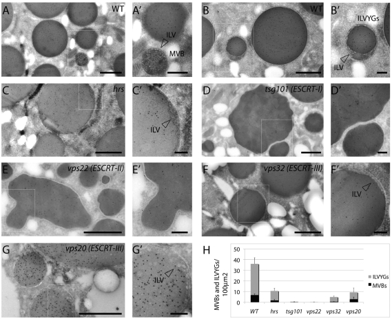
Ultrastructural characterization of ESCRT-mutants. ESCRT-I, ESCRT-II but not hrs or ESCRT-III are crucial for MVB biogenesis. (A-G) Yolk protein immunolabeling with 10 nm gold particles on cryosections of Drosophila oocytes. WT stage 9-10 oocytes show accumulation of yolk proteins in the yolk granules (A). Isolated MVBs containing ILVs are also frequently observed in the ooplasm (A′). Additionally ILV-containing yolk granules (ILVYGs), derived from fusion with MVBs, are frequently observed (B). Magnification (B′) shows that ILVYGs contain ILVs. (C) In hrs (ESCRT-0) mutants, there is a similar phenotype, with presence of isolated MVBs and ILVYGs, although they are less abundant than in WT oocytes (arrowhead in C′ indicates ILV). (D) In the tsg101 (ESCRT-I) mutants, the yolk granules still accumulate with large diameters; however, ILVYGs and isolated MVBs are absent. vps22 (ESCRT-II) mutant oocytes (E) also lack ILVYGs and isolated MVBs. (D′-E′) Magnifications show lack of ILVs. Compared with WT, the tsg101 and vps22 yolk granules are irregularly shaped. (F,G) By contrast, the vps32 and vps20 (ESCRT-III) mutant oocytes possess regularly shaped ILVYGs as well as isolated MVBs, which are more similar to hrs. Scale bars: 1 μm (A-G), 0.25 μm (A′-G′). (H) Quantification of the number of ILVYGs and MVBs per 100 μm2 in WT and mutant stage 10 ovaries. The number of ILVYGs and MVBs formed in the ESCRT-III mutants analyzed is similar to that of hrs mutant. By contrast, ESCRT-I and ESCRT-II mutant oocytes contains almost no ILVYGs and MVBs. Results are means ± s.d.

