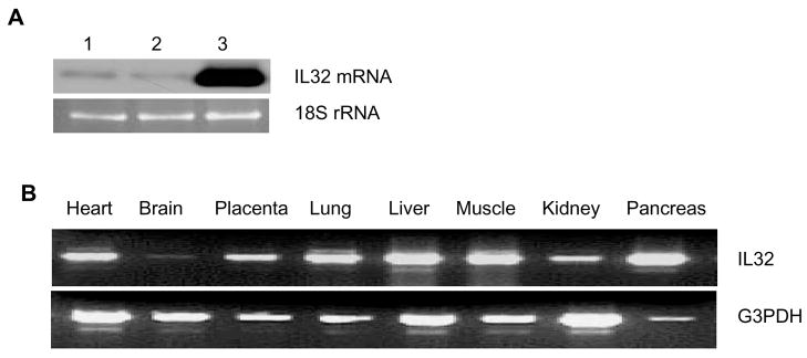
Figure 1. IL-32 is present in human endothelial cells and Akt induces its expression.
HUVECs were infected with control Ad β-gal (lane 2) or AdAkt (lane 3) adenoviral vector (MOI=10), respectively, for 48 hours. Uninfected cells were used as a control (lane 1). IL-32 mRNA was analyzed by Northern blot. 18S rRNA, visualized with ethidium bromide and UV, was used as a loading control (Panel A). IL-32 was amplified using a human tissue cDNA panel by PCR, and G3PDH was used as an internal control (Panel B). Each experiment was repeated at least twice. Representative images were shown.

