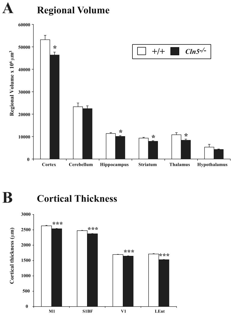
Figure 1.
Regional atrophy and cortical thinning in Cln5 deficient mice. (A) Unbiased Cavalieri estimates of regional volume revealed significant atrophy of the cortex, hippocampus, striatum and thalamus in 12 month old Cln5 deficient mice (Cln5-/-) vs. littermate controls (+/+). In contrast, the cerebellum and hypothalamus did not exhibit significant reduction in volume in mutant mice (mean±SEM; * p<0.05; ANOVA with post-hoc Bonferroni analysis). (B) Cortical thickness measurements of primary motor (M1), primary somatosensory (S1BF), primary visual (V1) and lateral entorhinal (LEnt) cortex revealed a significant thinning of these cortical regions in 12 month old Cln5-/- mice vs. littermate controls (mean±SEM; *** p<0.001; ANOVA with post-hoc Bonferroni analysis).

