Figure 4.
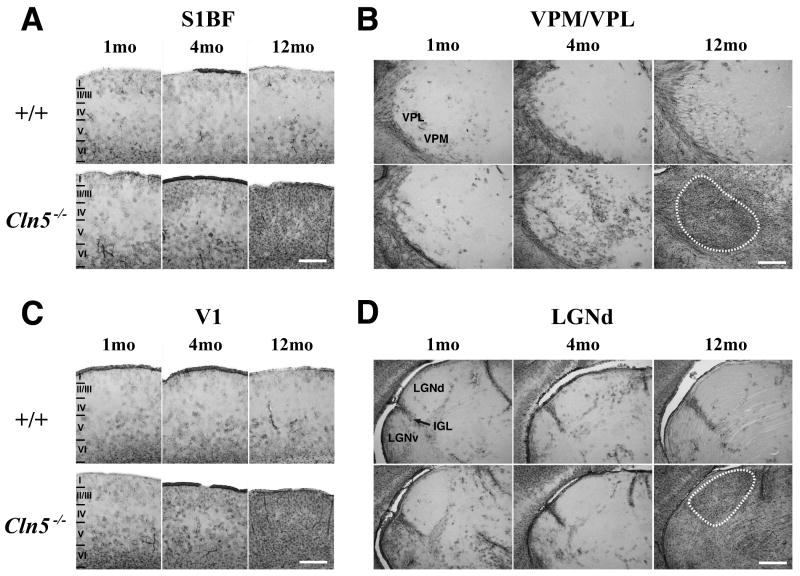
Progressive astrocytosis in the thalamocortical system of Cln5 deficient mice. (A-D) Immunohistochemical staining for glial fibrillary associated protein (GFAP) reveals the pronounced upregulation of this marker of astrocytosis with increased age in the somatosensory barrelfield cortex (S1BF, A) primary visual cortex (V1, C) of Cln5-/- mice compared to age matched controls (+/+). (A, C) At one month of age only few scattered GFAP immunoreactive astrocytes were evident mostly in deeper laminae of both S1BF and V1 in control mice, with similar staining evident in Cln5-/- mice. In these mutant mice at 4 months of age GFAP immunoreactivity became more pronounced in both deeper laminae (IV-VI) and superficial laminae (I-III) of both cortical regions, becoming markedly more intense and spreading to involve all laminae at 12 months of age. Laminar boundaries are indicated by roman numerals. (B, D) A similar progressive increase in the distribution and intensity of GFAP immunoreactivity was also evident in the ventral posterior (VPM/VPL, B) and dorsal lateral geniculate (LGNd, D) thalamic nucleus, which project to S1BF and V1, respectively. At 1 month of age GFAP immunoreactivity appeared similar in control and Cln5-/- mice, but numerous scattered GFAP-positive astrocytes were evident in VPM/VPL at 4 months of age. In contrast LGNd contained few GFAP immunoreactive astrocytes until 12 months of age when intense GFAP staining delineated both VPM/VPL and LGNd. The boundaries of these nuclei are indicated by white dashed lines. Scale bar = 300 μm.

