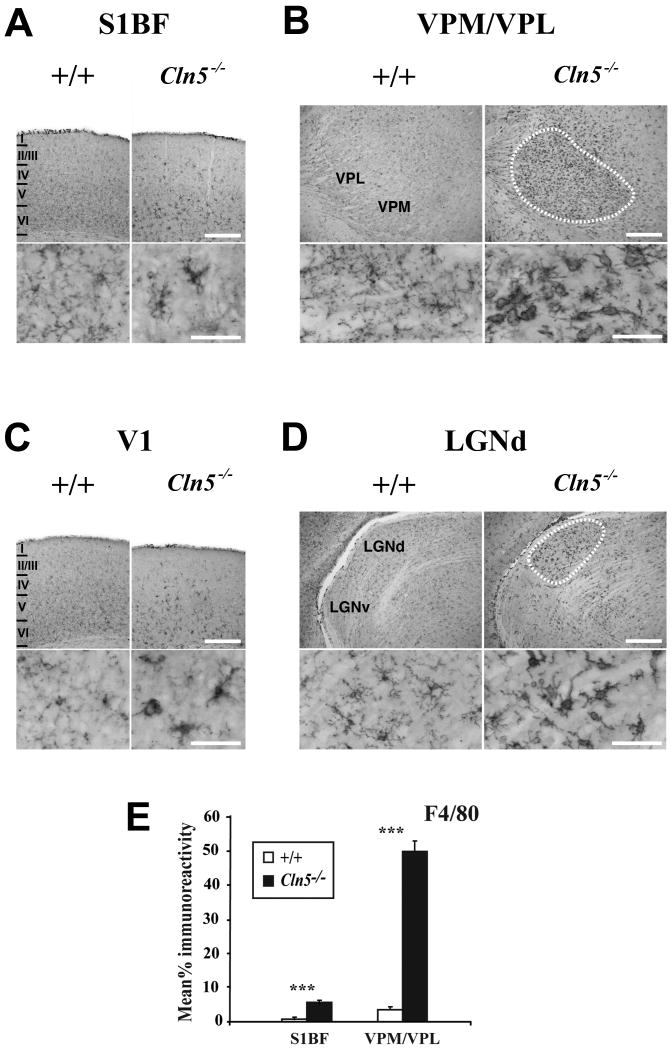
Figure 6.
Activation of microglia in 12 month old Cln5 deficient mice. (A, C) Immunohistochemical staining for the microglial marker F4/80 reveals activated microglia to be more frequent within the deeper laminae (IV-VI) of the somatosensory cortex (S1BF, A) and primary visual cortex (V1, B) of Cln5-/- mice at 12 months of age compared to age matched control mice (+/+). In mutant mice, microglia displayed more intense F4/80 immunoreactivity with enlarged soma and fewer ramified processes (inserts), although complete transformation to brain macrophage like morphology was rare. Laminar boundaries are indicated by roman numerals. (B, D) Localized microglial activation within individual thalamic nuclei of 12 month old Cln5-/- mice is also revealed by F4/80 immunoreactivity. In these mutant mice, intensely stained F4/80 positive microglia with brain macrophage like morphology were present in the ventral posterior (VPM/VPL), nucleus (B) and dorsal lateral geniculate nucleus (D), but were virtually absent from adjacent thalamic nuclei (including the ventral lateral geniculate nucleus, LGNv) and were not present in age matched controls. The boundaries of these nuclei are indicated by white dashed lines. Scale bar = 300 μm; 50 μm in inserts. (E) Thresholding image analysis confirms the progressive and significant increase in F4/80 staining in both S1BF and VPM/VPL of Cln5 deficient mice (Cln5-/-) with increased age, compared to age matched controls (+/+). (mean±SEM; *** p<0.001; ANOVA with post-hoc Bonferroni analysis).

