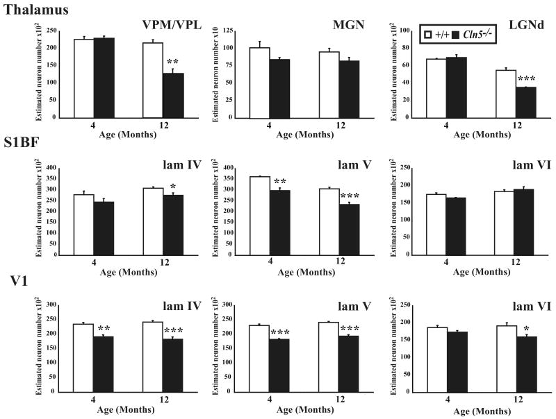
Figure 7.
Progressive loss of thalamic and cortical neurons in Cln5 deficient mice. (A) Histograms of unbiased optical fractionator estimates of the number of Nissl stained thalamic relay neurons in the ventral posterior (VPM/VPL), medial geniculate nucleus (MGN), and dorsal lateral geniculate nucleus (LGNd), of Cln5-/- mice and age-matched controls (+/+) at different stages of disease progression. The number of neurons in VPM/VPL and LGNd nuclei declined in Cln5-/- mice with increased age, with significant neuron loss evident in these nuclei in 12 month old mutant mice. In contrast no significant loss of MGN neurons was observed in Cln5-/- mice at either age. (B, C) Histograms of unbiased optical fractionator estimates of the number of Nissl stained lamina IV granule neurons, lamina V pyramidal neurons and lamina VI feedback neurons in the somatosensory cortex (S1BF, B) and primary visual cortex (V1, C) of Cln5-/- mice and age-matched controls (+/+) at different stages of disease progression. Both cortical regions displayed a progressive loss of cortical neurons in mutant mice, but this occurred earlier and in all laminae of V1. A significant loss of laminae IV and V neurons was already apparent in V1 of Cln5 deficient mice at 4 months of age, with additional significant neuron loss in lamina VI at 12 months of age. Neuron loss in S1BF progressed more slowly, but a significant loss of lamina V neurons was already evident in Cln5 deficient mice at 4 months of age, with an additional significant neuron loss in lamina IV of these mice at 12 months of age. (* p<0.05; ** p<0.01; *** p<0.001, ANOVA with post-hoc Bonferroni analysis).

